OVERVIEW
APPLICATIONS
INTERACTIVE APPLETS
HISTORY OF THE METHODS/FLOW CHART
PUBLICATIONS
EDUCATIONAL MATERIAL
ACKNOWLEDGEMENTS
ABOUT THE AUTHOR/CV
Copyright:
1996-2010
J.A. Sethian
Segmentation in Medical Imaging

|
|
|
|
|
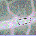
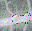
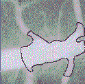
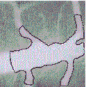
- When the curve passes over places where the image gradient (that is, the change in value from one pixel to the next) is small, we let the curve expand quickly.
- When the curve passes over places where the image gradient is large, we suspect we are near the boundary, and slow the curve down.
- In addition, we include a curvature term to the speed to add a little surface tension to the expanding contour.
|
|

|
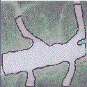
| Arterial Tree (1.3 MB) | Femurs and Surrounding Soft Tissue (232K) |

|

|
| Beating Heart(1.3 MB) | Reconstruction of Brain(232K) |
|
|
|
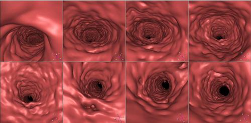
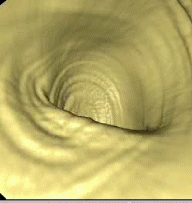
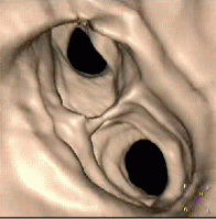
| Aortic Fly through | Virtual Endoscopy | Trachae Fly through |
Interactive Java Applet
Try out an interactive Java applet for automatic shape recovery
Advantages of this Approach
The level set approach allows the evolving front to change topology, break, and merge, which means that the evolving front can extract the boundaries of particularly intricate contours. In addition, the method works in three dimensions with almost no change, so three dimensional surfaces can be extracted as well.Details
The calculation was performed using a level set method to track the motion of an initial interface, which is embedded as the signed distance function. The resulting level set equation for front propagation is then updated using a first order in time, central difference in space scheme. The approach is similar to that of active contour models, in that a speed function is synthesized from the image gradient, and includes an expansive balloon force, a curvature-driven surface tension term, and Gaussian mollifier based on the local image-gradient. Additional moviesReferences
-
A Topology Independent Shape Modeling Scheme
,
Malladi, R., Sethian, J.A., and Vemuri, B.,
Proceedings of SPIE Conference on Geometric
Methods in Computer Vision II, Vol. 2031, San Diego, California,
pp. 246--258, July 1993.
List of downloadable publications -
Shape Modeling with Front Propagation: A Level Set Approach
,
Malladi, R., Sethian, J.A., and Vemuri, B.,
IEEE Trans. on Pattern Analysis and Machine Intelligence,
Vol. 17, No. 2, February 1995.
List of downloadable publications